In pathological studeis, in situ lesion detection and chemical composition analysis of histopathological specimen have long faced significant challenges due to labor-intensive pretreatment procedures and compromised sample integrity.
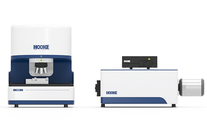
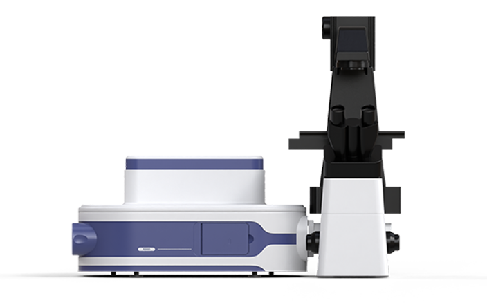
Hooke Instrument developed P300 Confocal Raman Spectrometer, characterized by its high signal-to-noise ratio, high stability, and high resolution, achieves label-free and damage-free examination of histopathological specimen, enabling precise analysis of spatial metabolic distribution.This provides a more intuitive detection method for rapid pathological examination and research into pathological and pharmacological mechanisms.The S3000 Ultrafast 3D fluorescence imaging system, leveraging its high-speed scanning and high-resolution confocal imaging features, demonstrates significant application value in research area such as pathology, drug efficacy analysis, and biomarker indentification.
-
Label-free Rapid Pathological Examination

-
Precise Categorization/Staging

-
Semi-quantitative Metabolic Analysis

-
Spatial-phenotype Research


Label-free Examination Approach Forhistopathological Specimen

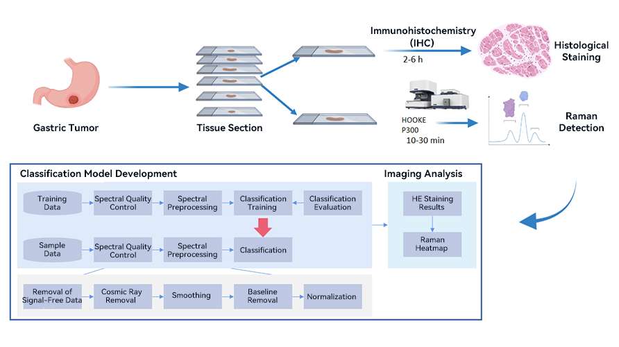
By comparing the differences in Raman spectra between abnormal tissues/cells (such as tumors) and normal tissues/cells, one can elucidate the pathological changes of molecular composition and structure alterations. This allows for the label-free analysis of different abnormal tissues and normal tissues, thereby facilitating the classification and identification of various sample types and their disease stages. This approach offers a phenotype-based examination strategy for reasech in tumor margin delineation, detection of distal invasive micro-foci, and tumor classification/grading.
Research on Pathological Mechanism
Confocal Raman spectroscopy analysis can elucidate the changes in cellular composition and metabolic patterns at the lesion site. The P300 Confocal Raman Spectrometer can rapidly and damage-freely identify the subtle biochemical changes in tissue and cellular metabolism resulting from pathological conditions. enabling semi-quantitative analysis. This capability allows for teh exploration of molecular mechanisms underlying pathological processes in different microenviroments. Meanwhile, the S3000 Ultra-fast 3D Fluorescence Imaging System serves as a highly efficient tool for evaluating biomarkers and assessing drug targeting.
Featured Products
-

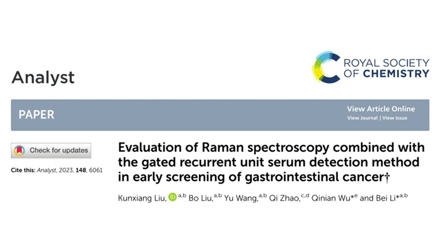 Analyst丨Evaluation of Raman spectroscopy combined with the gated recurrent unit serum detection method in early screening of gastrointestinal cancer2024.06.04
 -

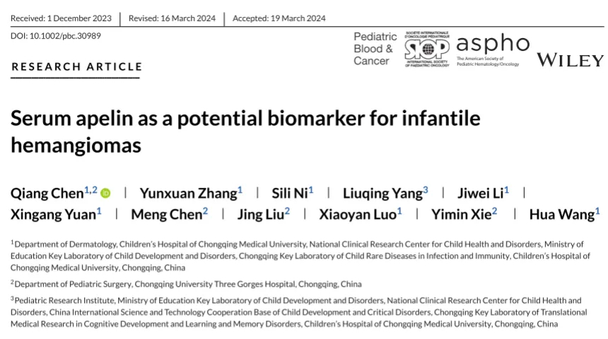 Pediatric blood & cancer丨Serum apelin as a potential biomarker for infantile hemangiomas2024.05.27
 -

 ACS Chemical Neuroscience丨In Vitro Diagnosis and Visualization of Cerebral Ischemia/Reperfusion Injury in Rats and Protective Effects of Ferulic Acid by Raman Biospectroscopy and Machine Learning2023.04.25
 -

 Frontiers in Cell and Developmental Biology丨Time-resolved extracellular matrix atlas of the developing human skin dermis2021.11.30
 -

 Advanced Healthcare Materials丨Unraveling an Innate Mechanism of Pathological Mineralization-Regulated Inflammation by a Nanobiomimetic System2021.10.13


