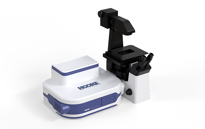
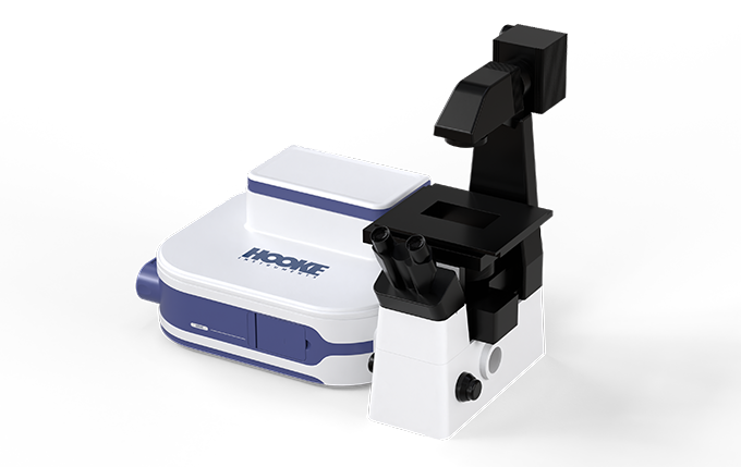
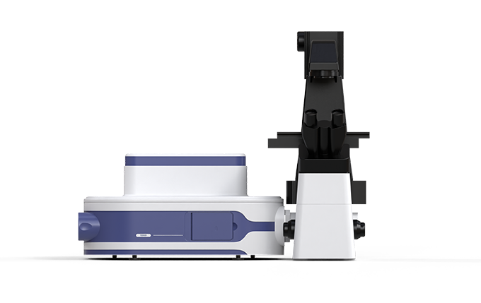
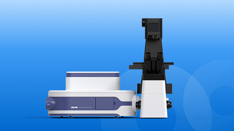
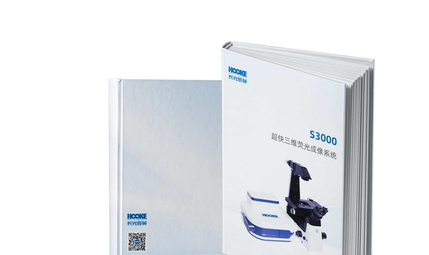
S3000
Ultrafast 3D Fluorescence Imaging System
Featuring a structured illumination spinning disk design with high optical transmittance, the system enables fluorescence confocal imaging using more convenient and longer-lasting non-laser light sources, overcoming the technical limitations of laser dependency. Within a compact setup, it minimizes optical path signal loss while achieving high-speed confocal imaging with superior weak-signal detection and imaging performance. The S3000 provides an innovative, time-saving, user-friendly, and reliable solution for 3D fluorescence tomography of cells, tissues, microscopic organisms, and microorganisms.
Brochures
Advantages
-
Laser-Independent
Instant Startup with No Waiting, Minimal Impact from Light Source Attenuation, and Extended Stable Operation Time.
-
Fast Array Imaging
Ultra-fast imaging with a minimum confocal exposure time of 20 ms per frame and a scanning refresh rate of up to 20/40 frames per second. Large-area stitching and multi-layer scanning are 10–20 times faster than point-scanning confocal microscopes. Real-time switching between confocal and wide-field modes allows for easy focal plane and target field selection.
-
Friendly for Biological Sample
Low phototoxicity and minimal bleaching, making it ideal for biological research.
-
Minimal Maintenance Required
No need to replace the light source throughout the instrument’s lifespan, reducing maintenance efforts and lowering operational costs.
-
Easy to Use
User-friendly software with an intuitive interface allows quick learning, enabling high-quality fluorescence data acquisition with half-day training.
-
Versatile Compatibility and Upgrades
Equipped with high-optical-quality microscopes and compatible with upgrades or integrations across popular microscope brands.
Working Principle
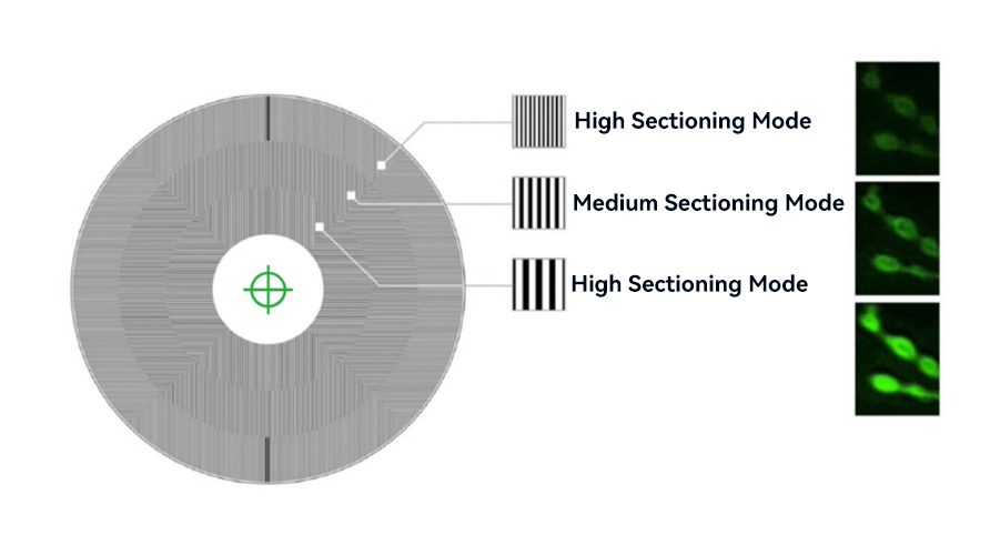
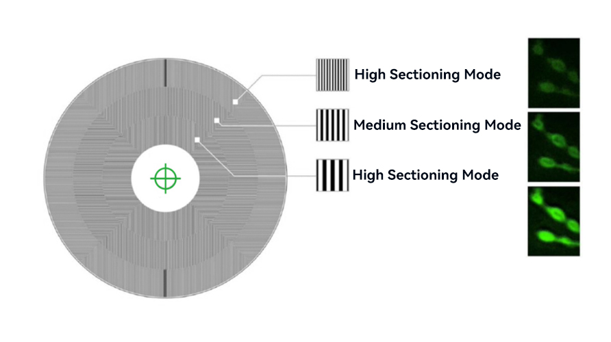
The confocal spinning disk rapidly rotates to scan and excite the sample using structured light modulated through slits. The compact design of the short optical path minimizes signal loss. Additionally, the high optical transmittance of the slit disk allows for the use of non-laser light sources in 3D fluorescence imaging.
Applications
-
 Cell Biology & Biochemistry
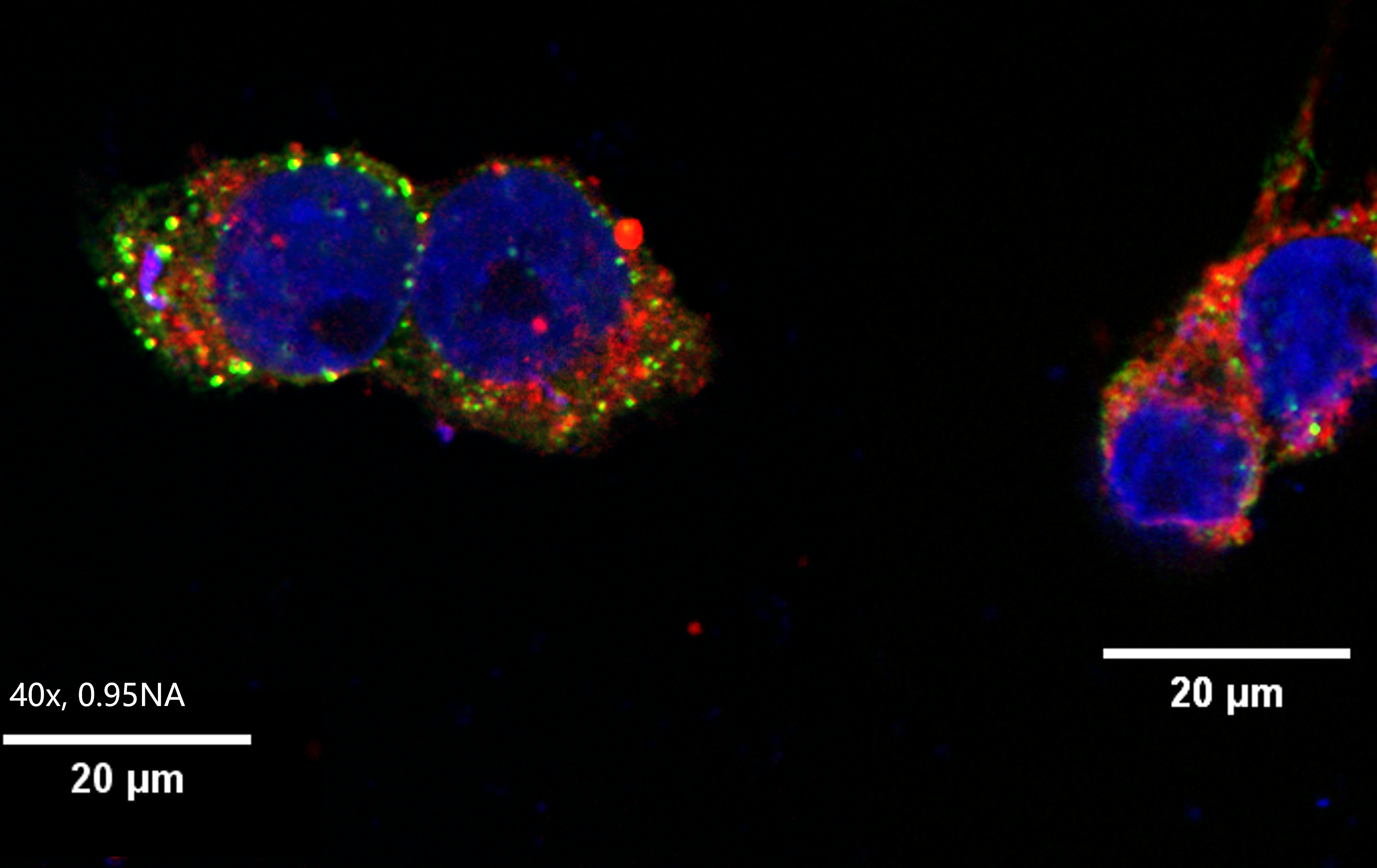
Capable of capturing cellular structures, cytoskeletal elements such as actin filaments and microtubules, cell membrane structures, receptors, organelle morphology and distribution changes, as well as apoptosis. Supports both fixed biological samples and live-cell dynamic imaging, enabling in-depth research and analysis based on these data.
-
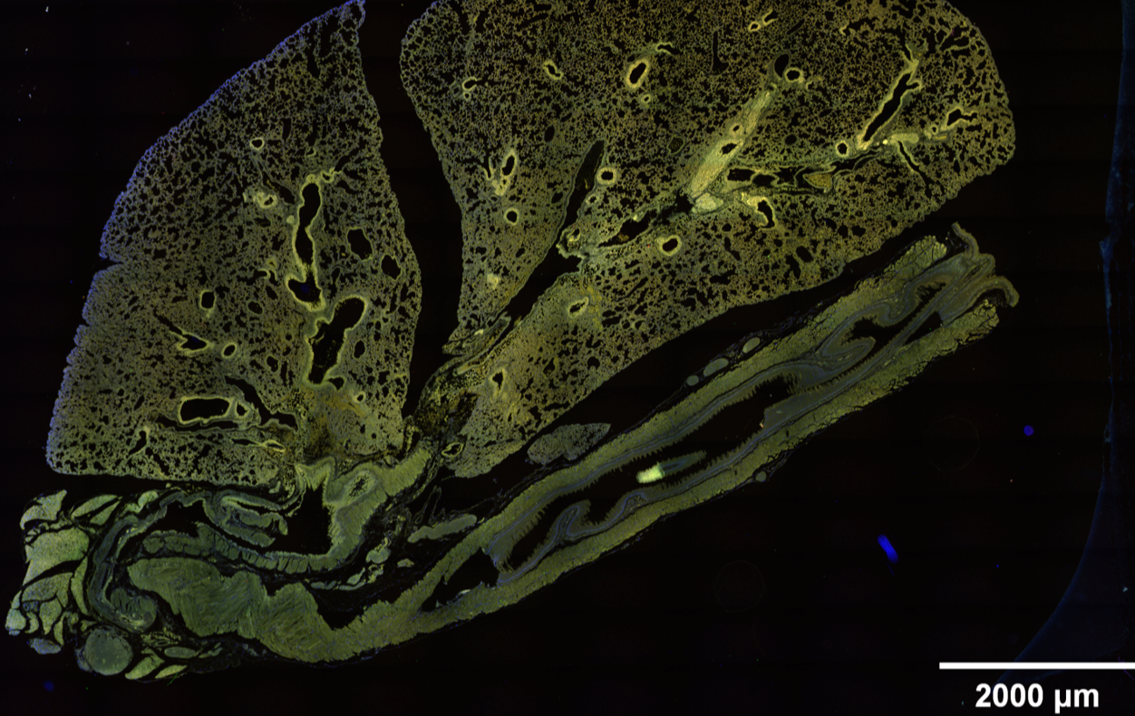 Animal & Plant Tissue Imaging
Supports multi-field stitching in the XY direction and multi-layer scanning in the Z direction, enabling rapid digital 3D imaging of sizable animal and plant tissue samples. Facilitates large-scale data acquisition and comprehensive fluorescence signal analysis for volumetric tissue studies.
-
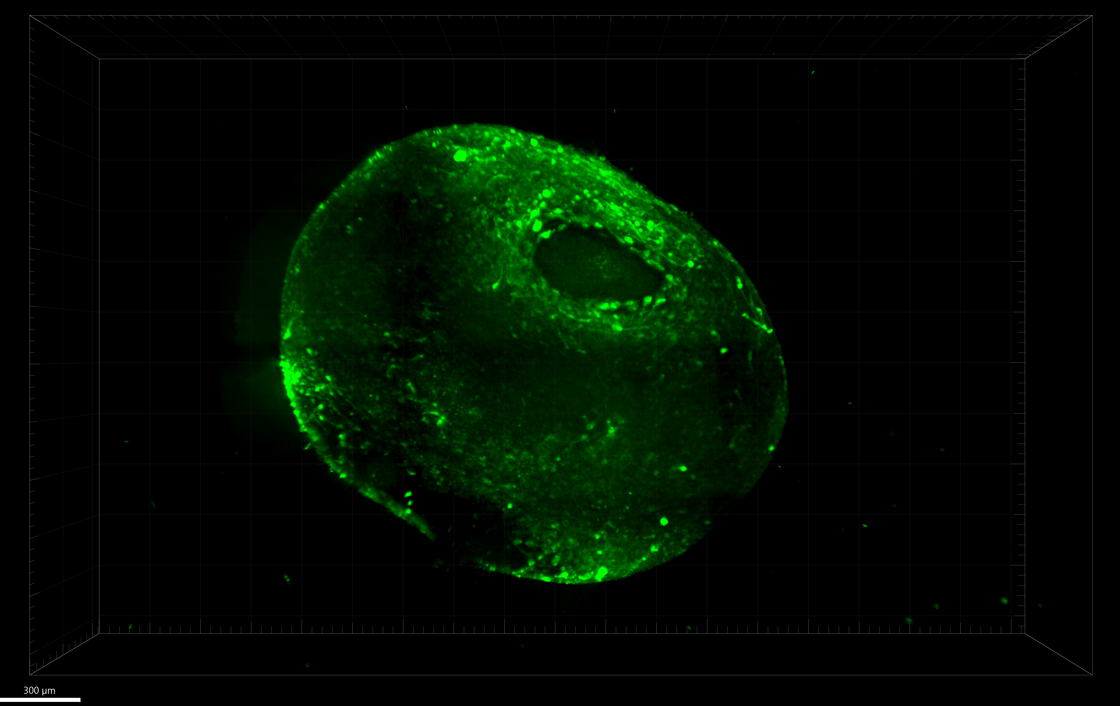 Organoid Research
Enables rapid,efficient scanning and 3D reconstruction of organoid structures. By integrating imaging data with organoid characteristics, it facilitates the study of drug efficacy and toxicity at the organoid level.
-
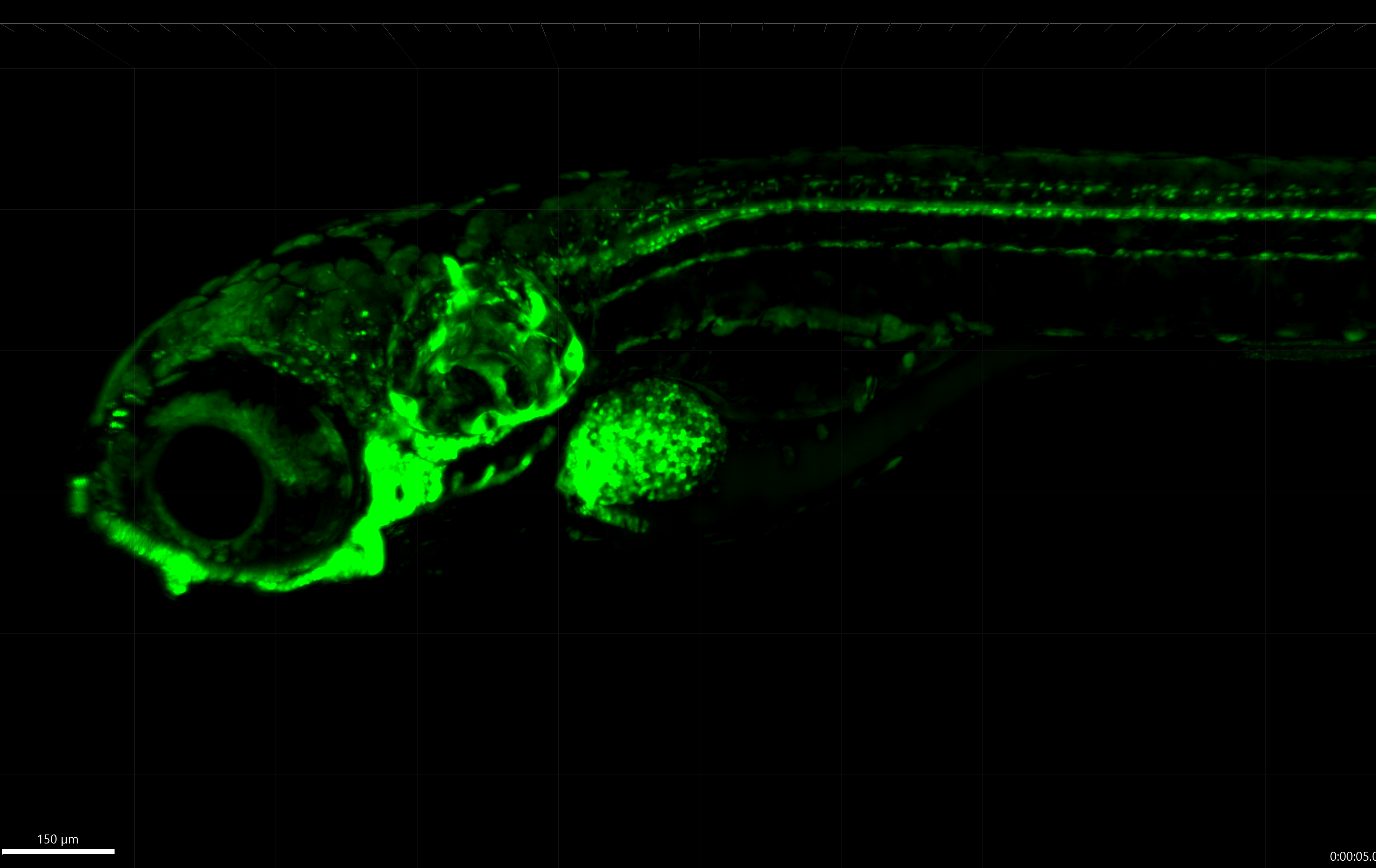 3D Imaging of Small Model Organisms (C. elegans, Zebrafish, etc.)
The system enables 3D imaging scans of fluorescent protein-labeled immune cells, neurons, vascular endothelial cells in zebrafish, as well as stained organs in zebrafish and C. elegans. This supports research in neuroscience, immunology, developmental biology, and toxicology.
-
 Cell Biology & Biochemistry
Capable of capturing cellular structures, cytoskeletal elements such as actin filaments and microtubules, cell membrane structures, receptors, organelle morphology and distribution changes, as well as apoptosis. Supports both fixed biological samples and live-cell dynamic imaging, enabling in-depth research and analysis based on these data.
-
 Animal & Plant Tissue Imaging
Supports multi-field stitching in the XY direction and multi-layer scanning in the Z direction, enabling rapid digital 3D imaging of sizable animal and plant tissue samples. Facilitates large-scale data acquisition and comprehensive fluorescence signal analysis for volumetric tissue studies.
-
 Organoid Research
Enables rapid,efficient scanning and 3D reconstruction of organoid structures. By integrating imaging data with organoid characteristics, it facilitates the study of drug efficacy and toxicity at the organoid level.
-
 3D Imaging of Small Model Organisms (C. elegans, Zebrafish, etc.)
The system enables 3D imaging scans of fluorescent protein-labeled immune cells, neurons, vascular endothelial cells in zebrafish, as well as stained organs in zebrafish and C. elegans. This supports research in neuroscience, immunology, developmental biology, and toxicology.
Publications
-

 J. Agric. Food. Chem.丨A Novel Endophytic Fungus Fusarium falciforme R‑423 for the Control of Rhizoctonia solani Root Rot in Pigeon Pea as Reflected by the Alleviation of Reactive xygen Species-Mediated Host Defense Responses2025.01.22
 -

 International Journal of Nanomedicine丨Combined ROS Sensitive Folate Receptor Targeted Micellar Formulations of Curcumin Effective Against Rheumatoid Arthritis in Rat Model2024.10.16
 -

 Pediatric blood & cancer丨Serum apelin as a potential biomarker for infantile hemangiomas2024.05.27
 -

 mLife丨Integrated identification of growth pattern and taxon of bacterium in gut microbiota via confocal fluorescence imaging‐oriented single‐cell sequencing2023.05.17
Configuration List
-
-
-
Filter Cube
-
Fluorescent Light Source
-
Back-illuminated sCMOS Camera
-
Motorized Microscope
-
DIC
-
Live cell incubator
-
-
hardware configuration
-
DAPI、GFP、FITC、YFP、DsRed、mCherry、CY5
-
Excitation wavelength 400-650 nm
Excitation wavelength365-770nm(NIR dye compatibility) -
20 fps
40 fps -
APO object lens
-
Compatible DIC imaging
-
PremixedC02: 5%
CO2:5-20%
-
-
-
Four filter cubes can be selected for one imaging
-
According to the characteristics of dyes
-
According to the experimental requirements
-
Support 4X-100X objective lens
-
According to the experimental requirements
-
According to the experimental requirements
View More


