Scatcher
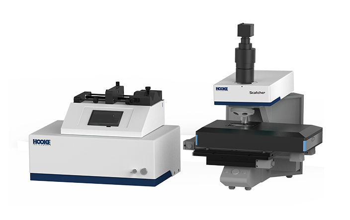
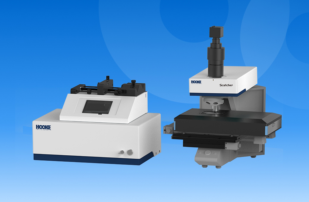
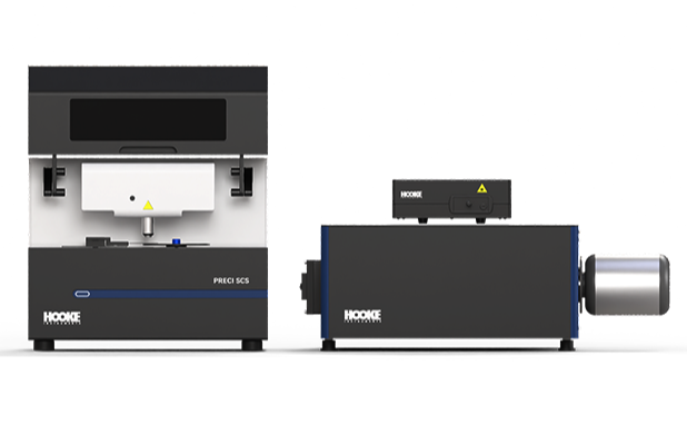
Single-Cell Optical Tweezers Sorter
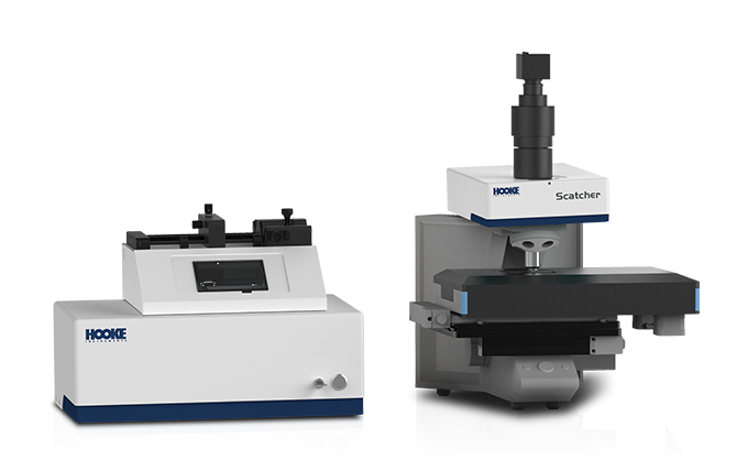
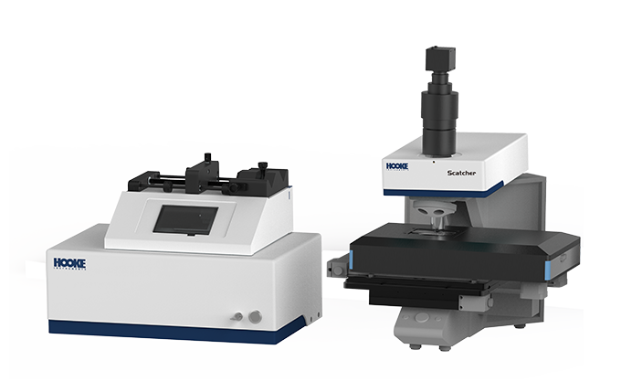
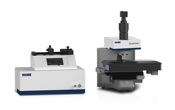
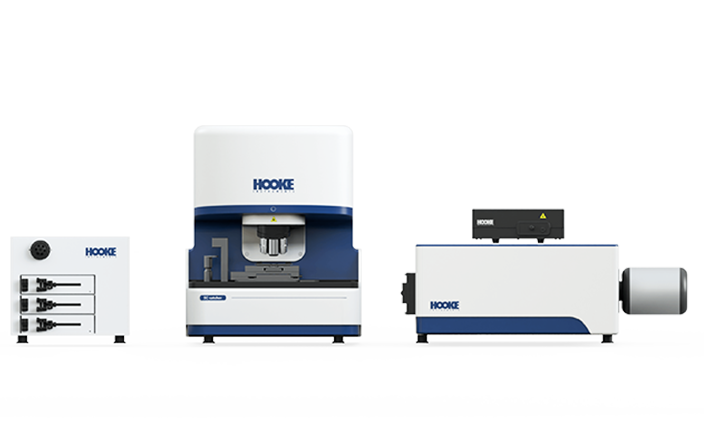
This system demonstrates robust micro-manipulation capabilities for micrometer-scale objects. Under a microscope, it enables efficient capture, controllable manipulation, and precise sorting of various types of cells and particles, providing a powerful tool for fields such as microbiology, drug development, and biomedical research.
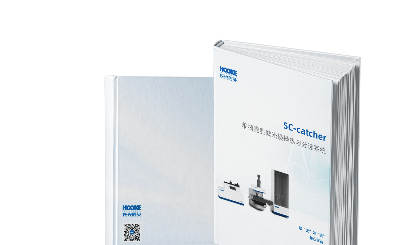
Brochures
Advantages
-
Ultra-High-Precision Micro-manipulation
Under the microscope, optical tweezers technology ensures high-precision capture and manipulation of single cells or particles, facilitating controlled movement and isolation with a sorting success rate of over 95%.
-
Ultra-High-Viability Single-Cell Sorting
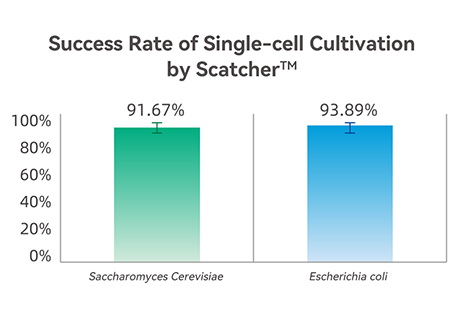
The system preserves the in situ state, growth activity, and metabolic functions of cells due to its low-damage feature, achieving a post-sorting culture success rate of over 90%.
-
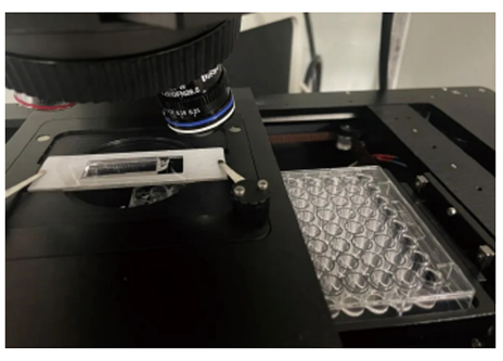
Automated Cell Receiving in Culture Plates
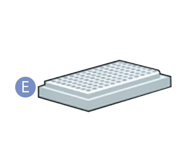
Equipped with a 96-well plate receiver module, the system enalbes the selection of specific cell reception areas on the plate as well as the number of cells to be received in each well based on experimental designs.
-
Modular design with Excellent Expansion Capability
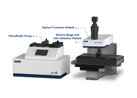
The optical tweezers module can be easily integrated into the user’s existing microscope, offering a cost-effective solution for upgrading microscope functionality.
-
Multiple Models to Meet Various Application Needs
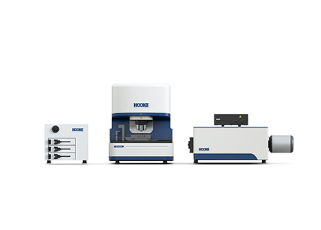
The instrument can be equipped with a fluorescence module and Raman spectroscope identification module, enabling multi-modal single-cell phenotypic detection and identification, thereby expanding its application scenarios. It can be installed in biosafety cabinets, anaerobic chambers and laminar flow hoods to better meet the needs of different applications.
-
Customized Microfluidic Cell Manipulation Chips
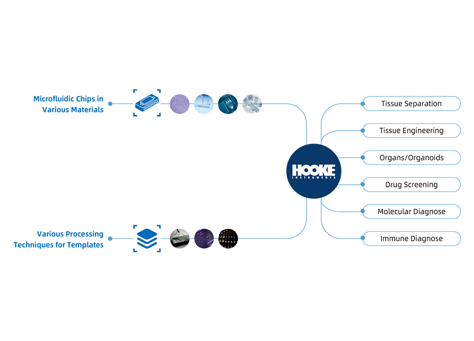
We can design and fabricate microfluidic chips tailored to users' specific experimental needs, offering comprehensive end-to-end solutions.
Working Principle
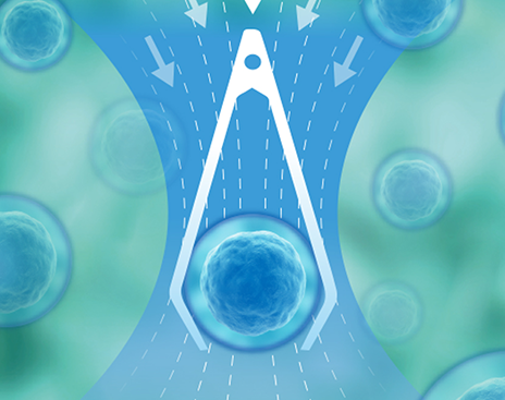
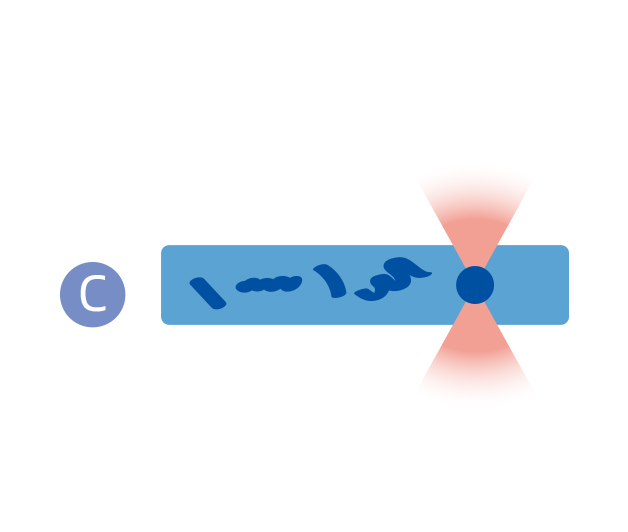
Optical tweezerss employ a highly concentrated laser beam to apply optical stresses to single cells, allowing them to be in situ, damage-free captured and manipulated. This approach enables researchers to achieve precise manipulation and isolation of single cells without disrupting cellular structure or compromising their functionality.


Workflow
-
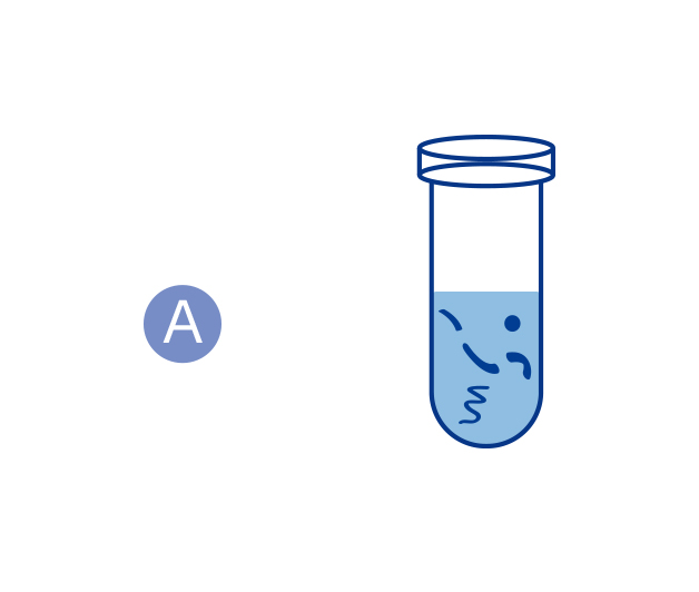
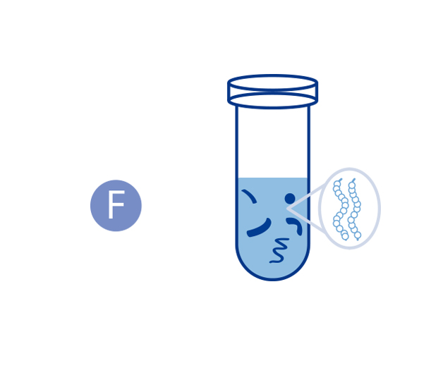
01Preparation of samples

-
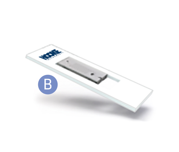
02On-chip sample injection

-
03Optical tweezers capture the target

-
04Cells of interest are encapsulated into microdroplests


-
05Cell collector

-
06Downstream experiments (sequencing,cultivation, etc.)

Applications
-
 Microbiology
Addressing the research challenge of uncultured microorganisms through a multidimensional and multiscale research platform.
-
 Medical Research
Phenotypic detection combined with single-cell analysis enhances detection speed and accuracy, driving advancements in medical research.
-
 Microbiology
Addressing the research challenge of uncultured microorganisms through a multidimensional and multiscale research platform.
-
 Medical Research
Phenotypic detection combined with single-cell analysis enhances detection speed and accuracy, driving advancements in medical research.
Publications
-

 Analytical Methods丨Rapid detection of beer spoilage bacteria based on label-free SERS technology2022.12.17
 -

 Talanta|Laser tweezers Raman spectroscopy combined with deep learning to classifymarine bacteria2022.03.29
Model Comparison
-
Functional Parameter
-
Applicable Sample Types
-
Sortable Cell Types
-
Sortable Cell Size
-
Single-cell Yield
-
Single-cell Viability Rate
-
Optical Tweezers Laser
-
Electric Stage
-
Cell Collector
-
Morphological Identification
-
Microscope
-
Fluorescence Recognition
-
Fluorescence Channel
-
Raman Spectral Identification
-
Confocal Raman Spectrometer
-
Data Analysis
-
-
-
-
Samples such as soil, marine, sediment, feces, pure cultures, etc
-
Bacteria, fungi, microalgae, mammalian cells, protoplasts, etc
-
0.5-20 μm
-
>95%
-
>90%
-
1064 ± 1 nm
-
Stage travel: X≥110 mm, Y≥75 mm
Repeatability:±1 μm
Knob control -
96-well plate collection
-
-
-
Bright field microscope
-
-
-
-
-
-
-
-
-
-
-
-
-
Samples such as soil, marine, sediment, feces, pure cultures, etc
-
Bacteria, fungi, microalgae, mammalian cells, protoplasts, etc
-
0.5-20 μm
-
>95%
-
>90%
-
1064 ± 1 nm
-
Stage travel: X≥110 mm, Y≥75 mm
Repeatability:±1 μm
Knob control -
8-tube strip collection
-
Bright field microscope
-
-
-
Spectral range:90-3600 cm-1
Spectral resolution:≤3 cm-1
SNR:≥30:1
Built-in multi-dimensional spectral calibration system -
Single-spectrum processing, Multi-spectrum batch processing, Characteristic peak analysis, Cluster analysis, Classification analysis, etc.
-
-
Samples such as soil, marine, sediment, feces, pure cultures, etc
-
Bacteria, fungi, microalgae, mammalian cells, protoplasts, etc
-
0.5-20 μm
-
>95%
-
>90%
-
1064 ± 1 nm
-
Stage travel: X≥110 mm, Y≥75 mm
Repeatability:±1 μm
Knob control -
96-well plate collection
-
Bright field microscope
-
4-channel fluorescence imaging: DAPI, GFP, Cy3, Cy5 (additional wavelengths customizable)
-
-
-
-
-
-
Samples such as soil, marine, sediment, feces, pure cultures, etc
-
Bacteria, fungi, microalgae, mammalian cells, protoplasts, etc
-
0.5-20 μm
-
>95%
-
>90%
-
1064 ± 1 nm
-
Opention
-
Opention
-
-
-
-
-
-
-
-
View More


