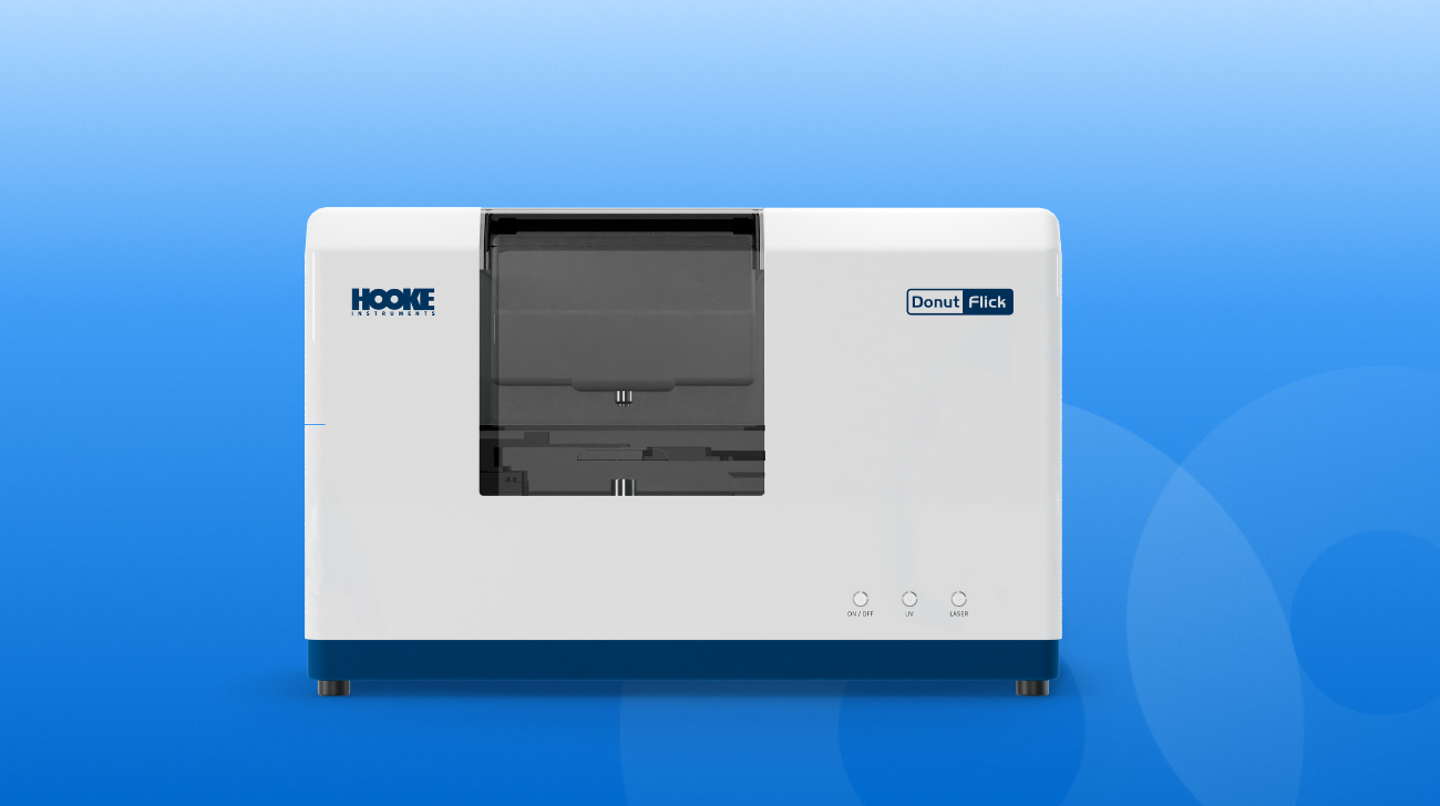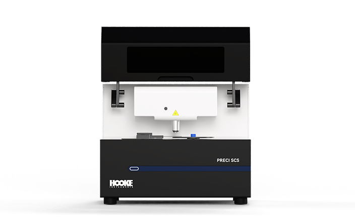


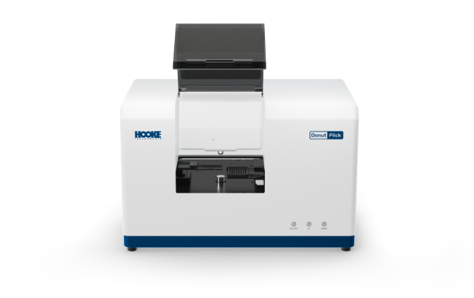
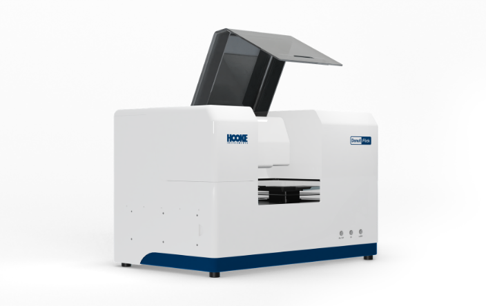
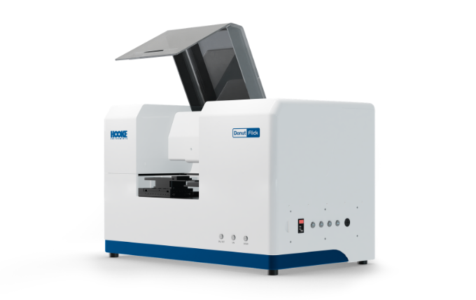
DonutFlick
Versatile Single-Cell Sorter
DonutFlick integrates laser-assisted gentle sorting with high-resolution microscopic imaging,enabling real-time visualization of cellular morphology and fluorescence signals during the sorting process. Whether working with valuable primary-cell suspensions or spatially resolved tissue sections, DonutFlick delivers precise single-cell isolation with recovery rate exceeding 95% while maintaining high cell viability. By preserving critical cellular information throughout the workflow, the system enables seamless integration with downstream sequencing and cell culture applications, bridging phenotype-based selection with genotype-driven analysis.
Brochures


Advantages
-
 Compatible with both cell suspensions and tissue sections: One System, Multiple Uses
Widely applicable to diverse sample types, including cell suspensions, formalin-fixed paraffin-embedded (FFPE) tissue sections, and cryosections, fully satisfying multidimensional single-cell research needs.
-
 High Single-Cell Recovery Rate & Preserved Cell Viability
Based on the principle of laser-induced forward transfer (LIFT), the single-cell recovery rate remains stable above 95%, and the cell survival/culture success rate exceeds 90%, providing high-quality samples for downstream investigation.
-
 New Strategies for Rare and Low-Abundance Cell Sorting
Ideal for low cell-density samples (as low as 10³ cells/mL) and rare cell populations (representing less than 0.1% of total samples), breaking through the limitations of conventional single-cell research.
-
 Visualized Precision Sorting with Full-Process Data Traceability
Equipped with a research-grade microscopic imaging system and a 4-channel fluorescence module, the entire sorting process is visually recorded. The image, coordinates, and fluorescence intensity of every single cell can be fully traced.
-
 Highly Automated and User-Friendly Operation
Features an integrated workflow covering wide-field scanning and image stitching, automatic focusing, AI-powered single-cell image recognition, one-click automated detection and isolation, and automated cell collection, significantly reducing experimental burdens and enhancing research efficiency.
-
 Seamless Integration with Downstream Multi-Omics Studies
Isolated cells can be directly utilized for single-clone culture, single-cell genomic amplification and sequencing, and single-cell proteomic analysis, effectively bridging phenotype-based selection with genotype-driven analysis.
Working Principle
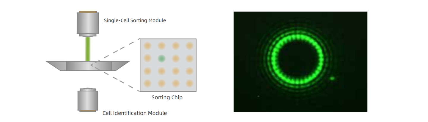
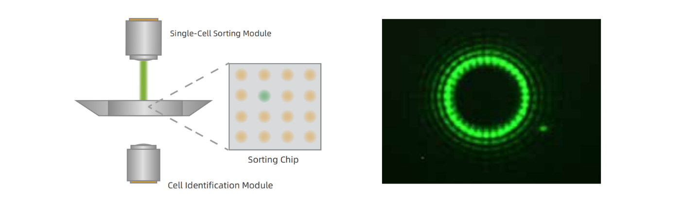
Utilizing Laser-Induced Forward Transfer (LIFT) technology combined with a beam shaping system to form a 'donut-shaped' laser beam, enabling precise non-contact separation of single cells and subcellular structures.
Applications
-
 Medical Research
Phenotypic detection combined with single-cell analysis enhances detection speed and accuracy, driving advancements in medical research.
-
 Biopharmaceuticals
From early-stage screening to formulation development, driving innovation in the biopharmaceutical field.
-
Medical Research
Phenotypic detection combined with single-cell analysis enhances detection speed and accuracy, driving advancements in medical research.
-
Biopharmaceuticals
From early-stage screening to formulation development, driving innovation in the biopharmaceutical field.
Product Parameters
-
Functional Indicator
-
Sortable cell types
-
Sortable cell size
-
Single-cell recovery rate
-
Cell culture success rate
-
Minimum sample concentration
-
Cell collection
-
Microscopic imaging
-
Intelligent recognition functions
-
Accessories
-
Downstream applications
-
-
Specification
-
Animal cells, tissue sections, plant cells, microorganisms, etc.
-
1-30 μm
-
≥95%
-
≥90%
-
1×10³ cells/mL
-
96-well plate, 8-strip microwell receiver
-
Research-grade brightfield imaging; 4-channel fluorescence imaging: DAPI, GFP, Cy3, Cy5; more wavelengths can be customized.
-
Morphological parameter analysis, fluorescence-intensity analysis and recognition, automatic numbering and localization.
-
Clean bench, microfluidic control device.
-
Cell culture, genomics, transcriptomics, proteomics, qPCR, etc.
-
View More