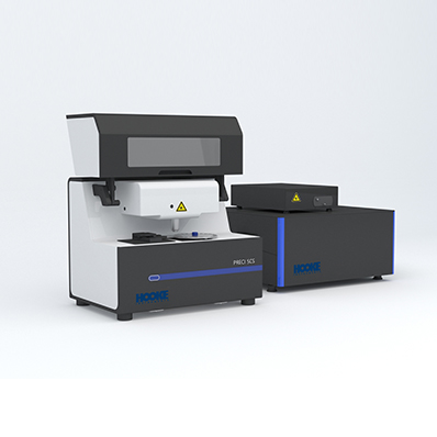
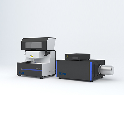
Ⅰ、Product introduction
PRECI SCS - R300 is a collection of confocal Raman spectroscopy detection system with single cell visual separation system for the integration of cell recognition and separation equipment, based on Raman spectroscopy identification of target bacteria, and sequencing on the single cell level, culture and other research, building the bridge unicellular phenotype and genotype, It provides a new strategy for single cell research in various fields. In addition, Raman separation technology can also be used for the identification and separation of microplastics and other small particles.
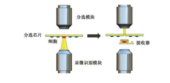
PRECI SCS-R300 single cell sorting principle
Ⅱ、Significance of microbial single cell research
Single cell research is a hot research field in biology. Over the past decade, important life processes such as cell differentiation, cell cycle and cellular immunity have been reunderstood in zoological studies using single-cell techniques. However, the research on microbial single cells is still in its infancy, so the realization of microbial single cell identification, rapid screening of functional bacteria, microbial identification, visual single cell isolation and high-throughput screening will provide technical breakthroughs for microbial research.

Traditional population research methods: the average value can not reflect the function of different cells in the community. Macrogene sequencing can reflect the composition and abundance of a community, but it still cannot pinpoint which microorganism plays a key role.
New technology for single-cell research: It can bypass the culture stage, establish the association between phenotype and genotype at the single-cell level, effectively solve the problems existing in population research, and provide new strategies for the study of functional microorganisms, mutant strains, drug-resistant bacteria, and unculture-resistant microorganisms.
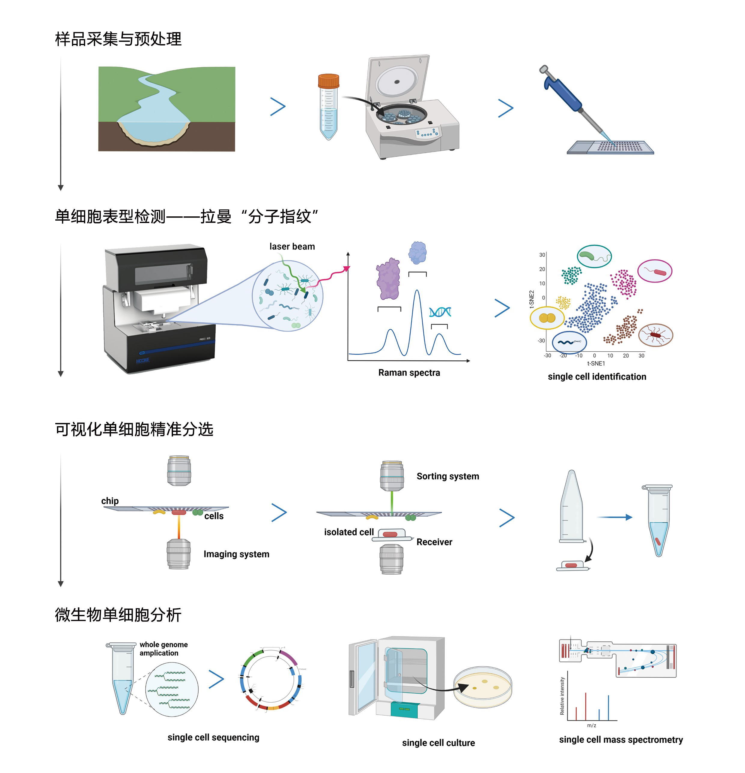
Ⅲ、Microbial single cell research strategies
Ⅳ、The research field
Cell biology (cell types identification, cell metabolism, etc.), plant developmental biology, genetics and breeding, genetic mechanism, etc.), genetics (stem cell growth, differentiation and mature dynamic monitoring, etc.), microbiology, microbial species identification and drug susceptibility testing, etc.), pathology (tumor precise separation, intraoperative navigation, fast pathologic analysis, etc.), pharmacokinetic (drugs Metabolic monitoring in cells, etc.), immunology (immune microenvironment, etc.), food science (food safety, fermentation engineering bacteria, etc.), etc.
Ⅴ、The experimental scheme
1. Single cell isolation of microorganisms based on morphology
According to the length, width, aspect ratio, regularity and other morphological indicators of the microbial cells, the identification, positioning and numbering are carried out, and the single cells of the target morphology are automatically separated.
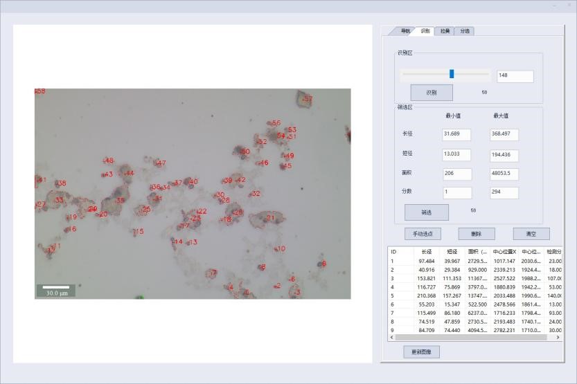
Microbial single cell intelligent image recognition function
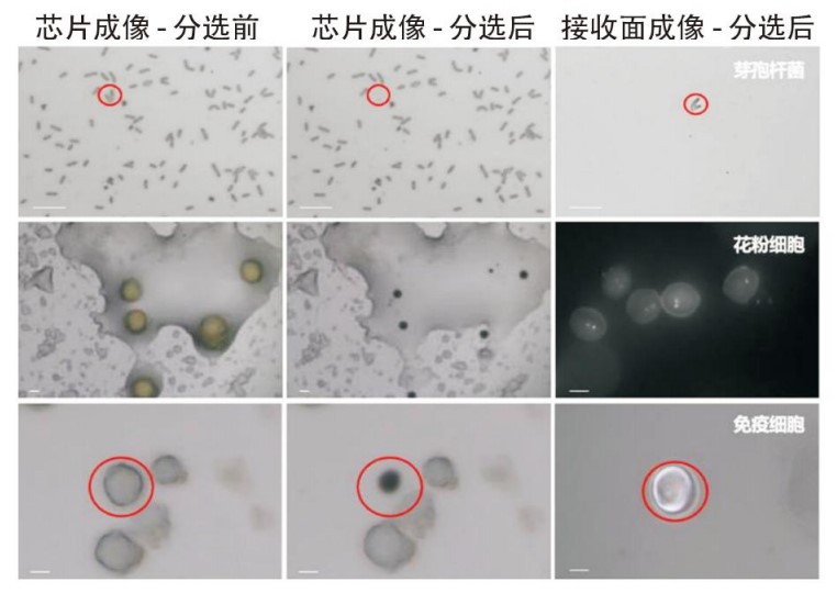
Single cell sorting results
2. Identification and isolation of microbial single cells based on Raman spectroscopy
2.1 Raman identification of drug-resistant bacteria/functional microorganisms (e.g. organic-degrading bacteria)
Heavy water labeling: for metabolically active microbial cells, D2O will enter the carbon skeleton through metabolism, form C-D bond, and make the peak position red shift to the silent zone. Using this method, the metabolic activity of bacteria/cells under stressful conditions (e.g., antibiotics, high temperature, high pressure, etc.) can be detected to identify drug-resistant bacteria or extremophiles.
13C and 15N stable isotope labeling: 13C and 15N isotope probes to the substrate complete tag for biomarkers, with a heavy isotope of biomarkers, the characteristics of Raman spectral lines with an effect will occur red mobile (isotope rate > 10%), using spectral analysis easily identified, and single target microorganisms.
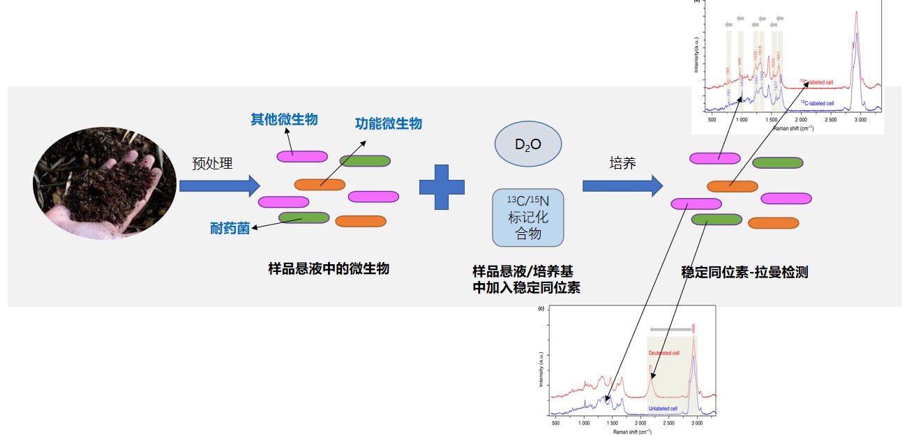
Raman identification process of functional bacteria
2.2 "Unknown bacteria" in the study sample (the species, function and other information of the target bacteria cannot be determined)
The "single cell Raman atlas" composed of the Raman signals of all compounds in a single cell of microorganism can be used as its "chemical fingerprint" to distinguish and identify cell species, which has the advantages of rapid, sensitive and non-destructive, and opens up a new field of microbial identification. Firstly, the cells were screened by morphological characteristics, and then the microorganisms in the samples were divided into different categories by Raman spectral clustering analysis. The microbial cells of each category were separated by visual ejection sorting, and the whole genome amplification and high-throughput sequencing analysis were performed.
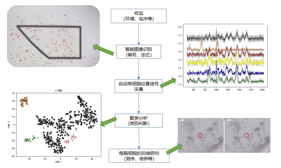
"Unknown Bacteria" single cell study protocol
3. Single cell isolation of microorganisms based on fluorescence
Single cells were identified and isolated based on the fluorescence signals of the target bacteria (autofluorescence, fluorescent dyes, probe labeling, FISH, etc.).
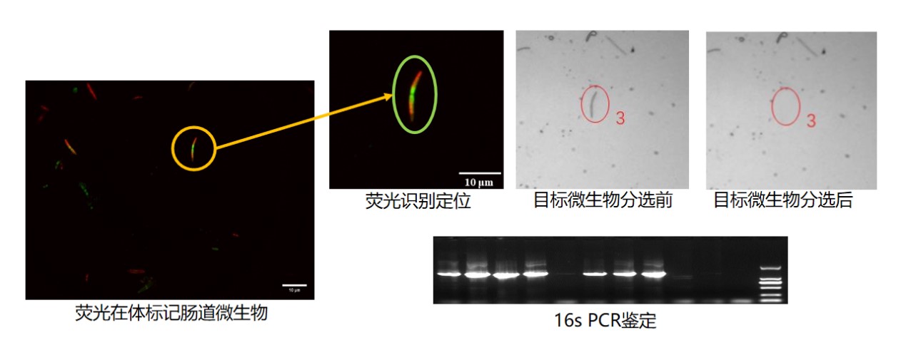
Single cell isolation of fluorescent bacteria
Ⅵ、The application case
1. Study on intestinal drug-resistant microorganisms
In this paper, a combination of isotope-Raman spectroscopy, single cell sorting and mini-Meta sequencing analysis was used to study the drug-resistant microbiota in human intestinal microbiota, and to reveal the correlation between intestinal drug-resistant bacteria and individual drug history.

The literature:Raman-activated sorting of antibiotic-resistant bacteria in human gut microbiota
2.D2O probe traced the horizontal transfer of drug resistance genes
Single-cell Raman spectroscopy combined with reverse D2O labeling (Raman-rD2O) is a sensitive and rapid phenotypic tool that can be used to track the spread of plasmids carrying antibiotic resistance genes (ARGs) from soil to clinical bacteria through transformation. Raman-rd2o sensitively identifies low abundance phenotypic resistant transformants from a large number of recipient cells, and then combines single cell sorting to obtain target transformants and perform gene identification to explore horizontal transfer of drug resistance genes.
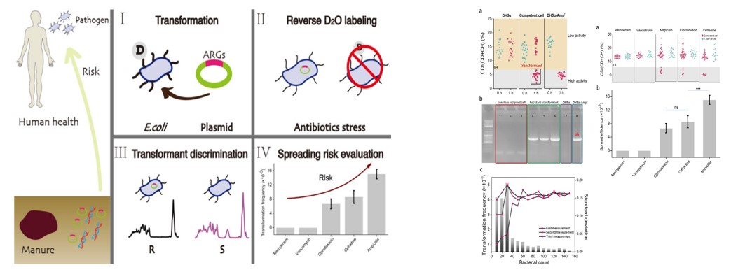
The literature:Phenotypic Tracking of Antibiotic Resistance Spread via Transformation for Environment to Clinic
3. Mini-metagenome analysis of thermophilic electroactive microorganisms
Raman single cell sorting, hierarchical clustering analysis and metagenomic sequencing were used to study the genes and metabolic functions of psychrophila.

The literature:Mini-metagenome analysis of psychrophilic electroactive biofilms based on single cell sorting
4. Fluorescence recognition and single cell isolation and culture of iron-reducing bacteria
In this paper, a Fe2+ specific fluorescent chemical probe (FSFC) was synthesized to identify and isolate ferric reducing bacteria (FeRM) from the community by using fluorescence detection and single cell sorting technology, and single cell culture was realized, and functional strains were successfully obtained.

The literature:Visualizing and isolating iron-reducing microorganisms at single cell level
5. Screening of engineered bacteria
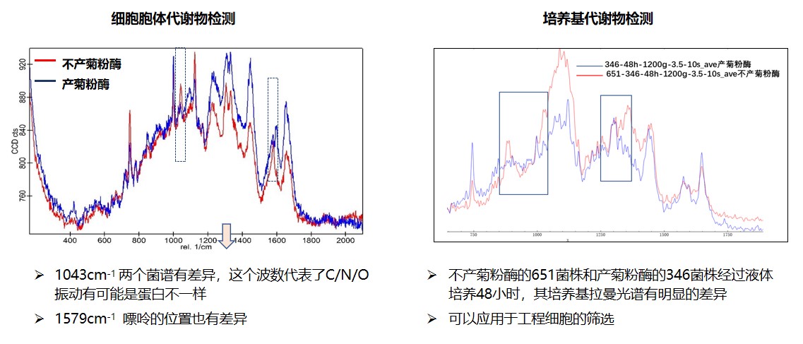
Raman technology was used to detect the cell body and medium metabolites of inulinase-producing yeast and control samples, and the differences in the Raman spectra of the engineered bacteria were determined. The database constructed by the database combined with visual single cell sorting culture was used to improve the screening efficiency of the engineered bacteria.
